Elekta Intraoperative Periscopic Viewer

- Maximize
Elekta Intraoperative Periscopic Viewer
Item Number :
1100-05-40
1100-05-40
Elekta Intraoperative Periscopic Viewer
 View PDF Catalog Closeout Items
Elekta Intraoperative Periscopic Viewer
Elekta Intraoperative Periscopic Viewer
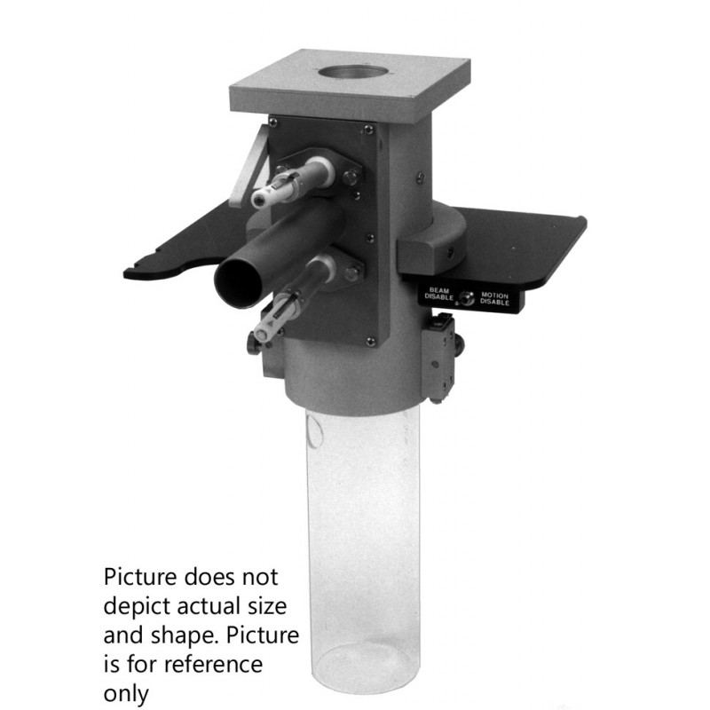
The periscopic viewer is used with the periscopic electron cones (Item 1100EC-1.9 to 1100EC-9.5) for intraoperative or intracavitary treatment. The periscopic viewer is made of stainless steel and clear anodized aluminum parts.
The most important feature of the periscopic viewer is the lateral docking. The bottom part of the barrel has a hinged door which opens to allow for the lateral docking of the periscopic electron cone into the viewer. The door has to be closed and secured to keep the periscopic electron cone in alignment with the beam. The periscopic electron cone can be held in place by the stainless steel locking knob.
If necessary the periscopic electron cone can slide approximately 8” into the barrel of the periscopic viewer. If the periscopic electron cone is being held in place by the locking knob this will have to be loosened to allow the retraction of the periscopic electron cone into the barrel.
Centering washers, four screws and beveled washers are used to attach the periscopic viewer to the plate that slides into the electron slot of the treatment machine. To adjust the periscopic viewer to the central axis of the beam, remove centering washers, position the periscopic viewer to central axis and using the beveled washers, tighten the screws. The centering washers keep the periscopic viewer centered to the plate.
Inside the periscopic viewer is a highly polished stainless steel mirror. The mirror is held and retracted by two independent springs. This prevents the possibility of the mirror remaining in the field if a spring should fail. A lever on the outside of the viewer adjusts the angle of the mirror for viewing through the periscopic electron cone.
At the top of the barrel of the periscopic viewer a sheet of 0.002" thick Mylar® is secured to prevent any foreign matter from entering the treatment field.
On the outside of the periscopic viewer are two pen light holders which are mounted on swivel sockets. The pen lights can be adjusted to allow maximum light into the periscopic viewer and electron cone. The pen light holders can be removed to allow for a fiber optics light source. A light source can also be inserted into the 2.5 cm hole in the side of periscopic electron cones that are 3.8 cm in diameter or larger. Ceiling spotlights or flashlights can be aimed at the periscopic electron cone to transmit light to the treatment area. Located between the pen holders is the periscopic viewer tube for viewing.
Custom electronics may need to be supplied by the accelerator manufacturer. RPD will assist the customer in determining what is needed for electronics.
As a safety precaution the gantry rotation power and couch vertical drive power should be disabled or turned off just before the lateral docking into the periscopic viewer. This prevents any accidental movement of the gantry or couch.
Sterilization: Gas - Sterrad NX-100


